Imaging-based screening for anti-cancer drug discovery
Cancer is the most common cause of cancer in the Western world. While in the past two decades understanding of mechanisms of cancer development and progression has resulted in various new targeted cancer therapeutics that entered the clinic, the overall success rate of novel candidate anticancer drugs that make it to the clinic is low. This is largely due to limited efficacy in the clinical setting. There is an urgent need to better select drug leads that can progress to the clinic.
To bridge this need the past few years have seen a strong push for the development of patient-derived 3D cancer organoid models that can be applied for drug screening. So far these 3D cancer organoid screens are largely based on simple cell survival measurements and lack quantitative biomarkers that reflect key elements of cancer development and progression. Our hypothesis is that integration of such biomarkers in patient-derived 3D cancer organoid models would further enable the early selection of the right candidate drugs for further drug development.
The aim of this project is to establish fluorescent protein biomarker 3D cancer test systems and optimize these models for drug screening application using high throughput microscopy approaches. We will integrate a panel of relevant fluorescent markers in breast, colon and liver cancer 3D models. We will next optimize these models for high content imaging approaches and then establish image analysis pipelines to extract quantitative biomarker activity at the single cell level in space and time. Finally we will perform a small proof-of-concept demonstration screen with a selected set of targeted anticancer drugs to establish the applicability of our novel fluorescent protein biomarker 3D cancer models for future commercial screening campaigns.
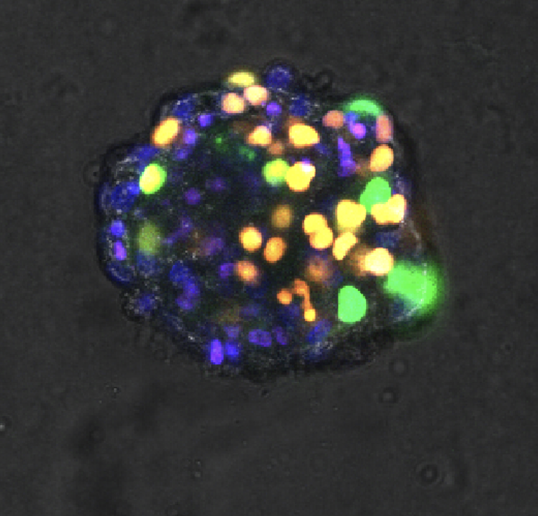
Legend: breast cancer microtissue expressing the FUCCI reporter, cultured in a 3D environment and imaged with confocal microscopy.
