Measuring stroke treatment effect using images acquired during treatment
Ischemic stroke is a worldwide major cause of death and permanent disability. In the Netherlands more than 26,000 ischemic stroke patients are admitted to a hospital per year. A recent breakthrough study showed that patient outcome in acute stroke caused by an occlusion of a large vessel could be improved with endovascular therapy. However, not all patients benefit from this intervention, and the current score to measure treatment success (TICI) measure recanalization, and does not match well with outcome. This project therefore aims to develop methods to quantify and assess reperfusion of small vessels (based on images during the intervention) as a treatment outcome.
To this end, Philips Healthcare will collaborate with the Interventional Neuroradiology Section and the Biomedical Imaging Group Rotterdam (both Erasmus MC) to address this challenge. Philips Healthcare is one of the largest players in interventional imaging systems, with great expertise in medical imaging. The Section of Interventional Neuroradiology is one of the largest in The Netherlands(part of the Erasmus MC Stroke Center ), and has world-class expertise in treating stroke patients. The BIGR group, with great expertise in medical image processing, completes the expertise required to address this challenge.
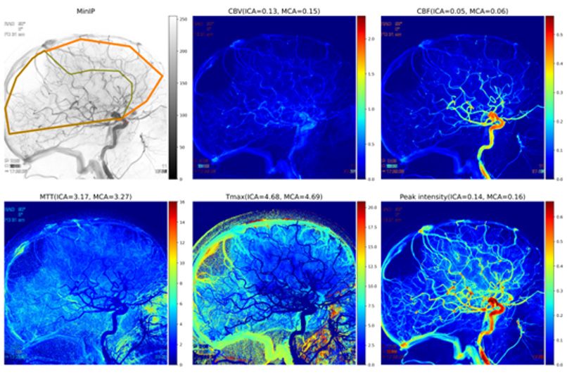
In our approach, the replacement of the recanalization concept, the current measure of treatment success, with a measure of reperfusion is a major innovation. To this end, we developed and evaluated methods to quantitatively assess reperfusion in EVT procedures, both in the traditional way (autoTICI) and using novel quantitative DSA perfusion techniques. We demonstrated that autoTICI strongly correlates to the manual scoring, and can also replace manual scoring in outcome prediction models. In addition, preliminary studies in data acquired in the MR Clean trials suggest that there are perfusion parameters that may discriminate patients with good and bad outcome, in case the recanalization was scored as good. The project results may help clinicians to select patients for additional pharmacological treatment in case of insufficient reperfusion after treatment. This may lead to improved patient outcome in stroke treatment. Per-procedural (in the angiosuite) identification of the absence of reperfusion of brain tissue by DSA image analysis is therefore of great importance for the patient and society. As a first step, we are currently assessing the autoTICI scoring system in the Erasmus MC. In addition, Philips and Erasmus MC, together with Erasmus University, are continuing their collaboration in the newly established ICAI Stroke Lab.


